Research


Our cancer research is focused on G proteins, GPCRs, HNSCC, and precision immunotherapy. Explore the panels below to learn more about our work.


G Proteins / GPCRS and Cancer

GPCRs, the largest family of cell-surface molecules involved in signal transmission, have recently emerged as crucial players in tumour growth, angiogenesis, immune evasion, and metastasis.

HNSCC / ORAL CANCER / PI3K-MTOR and Hippo Pathways

The PI3K/mTOR and Hippo signaling pathways are the most frequently dysregulated signaling mechanisms in HNSCC. mTOR inhibition halts tumor progression, and the Hippo pathway has recently emerged as a novel therapeutic target.

IMMUNE ONCOLOGY

The advent of the immunotherapy era based on the success of immune checkpoint blockade (ICB) has revolutionized cancer treatment. We are developing new combination therapies targeting cancer-specific immune evasive mechanisms and immune GPCRs to increase the response to ICB, aiming at achieving durable cancer remission (cure).

UVEAL MELANOMA

Around 90% of ocular melanomas harbor gain-of- function mutations in GNAQ or GNA11, where they act as driver oncogenes. We are developing new signal transduction-based multimodal precision therapies for primary and metastatic uveal melanoma.
Oncogenic signaling by G proteins and GPCRs

Our laboratory pioneered the study of the oncogenic activity of G proteins and G protein coupled receptors (GPCRs), including virally-encoded GPCRs, and how their signaling circuitry regulates normal and aberrant cell growth and metastasis. We demonstrated the potent transforming activity of G proteins and GPCRs, including Gαq (GNAQ), Gα12 (GNA12) and their coupled GPCRs, and showed that this remarkable biological activity requires a novel signaling network involving mitogen activated kinases (MAPK), ERK, JNK, p38, and their regulation by small GTPases of the Ras and Rho family. The latter had broad implications in the signal transduction field, as we showed that Rho GTPases, which were known for their cytoskeletal effects, stimulate JNK, thereby providing the first demonstration that Rho proteins are integral components of signaling routes linking the cell surface to the nucleus. We also made the unexpected discovery that rather than G protein α subunits (Gα), Gβγ subunits initiate the activate ERK through Ras, thus providing the first demonstration that cell growth promotion by GPCRs involves a complex protein-protein interaction network rather than relying solely on diffusible second messengers. We also reported the first studies supporting that non-receptor tyrosine kinases and tyrosine kinase growth factor receptors converge with GPCRs in the activation of Rho-regulated MAPK signaling networks to control the expression of growth promoting genes.


GPCR initiates Kaposi's sarcomagenesis

Our team revealed the transforming potential of a GPCR encoded by the open reading frame 74 of the Kaposi’s sarcoma (KS)-associated herpesvirus (KSHV) genome, thus providing a direct link between GPCRs and human viral-associated malignancies. For these studies, we developed a novel high throughput animal model system enabling the tissue-specific viral gene delivery and expression in mouse endothelial cells in vivo. This approach revealed that among all candidate KSHV oncogenes, only the viral GPCR was able to recapitulate KS sarcoma pathogenesis. Dissection of the underlying mechanism led to the discovery that the stimulation of AKT and mTOR by the viral GPCR were strictly necessary for sarcomagenesis. Our basic studies provided a rationale for the clinical evaluation by other groups of rapamycin, an mTOR inhibitor, in renal transplanted patients developing KS, which represented the first successful use of mTOR inhibiting agents to treat human malignancies. We also showed that the GPCR signaling specificity can be exploited by targeting selectively the PI3Kγ isoform for KS treatment, thereby circumventing the potential immunosuppressive activity of mTOR inhibitors in AIDS patients, who are at high risk of developing KS.


G proteins and GPCRs in human malignancies

G protein-coupled receptors (GPCRs) represent the largest family of cell surface proteins involved in signal transmission. These receptors play key physiological roles, and their dysfunction contributes to some of the most prevalent human diseases. However, the contribution of GPCRs, their linked heterotrimeric G proteins, and their regulated signaling circuitries in cancer initiation, progression, immune evasion, metastatic spread, and cancer therapy resistance has not been thoroughly investigated, Emerging evidence from our laboratories at UCSD has revealed that malignant cells often hijack the normal physiological functions of GPCRs to proliferate autonomously, evade immune detection, enhance their nutrient and oxygen supply, invade their surrounding tissues, and disseminate to other organs. Strikingly, our recent analysis of human cancer genomes revealed an unanticipated high frequency of mutations in G proteins and GPCRs in most tumor types, which we refer to as the onco-GPCRome. Indeed, nearly 30% of human cancers harbor mutations in GPCRs or G proteins. For example, mutually exclusive activating mutations in GNAQ or GNA11 (encoding Gαq family members) occur in ~90% and ~4% of melanomas arising in the eye and skin, respectively, where they act as driver oncogenes. Remarkably, approximately 5% of all sequenced tumors harbor activated GNAS mutants, encoding a constitutively active Gαs. Oncogenic GNAS mutations were initially discovered in multiple endocrine tumors, such as growth hormone-secreting pituitary (28%) and thyroid adenomas (5%). However, we have recently found that GNAS is mutated in many human cancers, including colorectal carcinoma (CRC) (4%), pancreatic tumors (12%), hepatocellular carcinoma (2%) and, strikingly, appendix cancers (70%). In this regard, there is substantial epidemiological evidence supporting the efficacy of aspirin and other nonsteroidal anti-inflammatory drugs (NSAIDs) that block COX2 for the prevention of CRC. Our team discovered that prostaglandin E2 (PGE2), which accumulates due to COX2 overexpression, can act on CRC cells directly by stimulating the β-catenin pathway through Gs-linked PGE2 receptors, supporting the direct relevance of GNAS and its activated pathways for CRC initiation and progression. Overexpression of GPCRs and their activation by locally released and circulating agonists in an autocrine and paracrine fashion is also a frequent event in cancer. Overall, as GPCRs represent the target of >30% of all drugs in the market, we hypothesize that interfering with the human onco-GPCRome by targeting GPCRs, G proteins, or their downstream signaling pathways may provide a unique opportunity for the development of novel, mechanism-based strategies for cancer diagnosis, prevention and treatment. See also our current efforts targeting GNAQ in uveal melanoma, as well as harnessing the power of computational biology approaches to target tumor-immune GPCR as part of combination therapies with immune checkpoint blockade.

PI3K/AKT/mTOR activation and Hippo pathway dysregulation in oral cancer

While at the NIH, our team I had the opportunity to develop and lead a new research program on HNSCC, a disease that results in >300,000 deaths each year worldwide. Our laboratory made the seminal finding that the persistent activation of the PI3K/AKT/mTOR signaling pathway is the most frequently dysregulated molecular signaling mechanisms in HNSCC, including tobacco-related and human papilloma virus (HPV)-associated lesions (>80% of all HPV- and HPV+ cases).  Furthermore, through the development and use of a large series of genetically engineered mouse models (GEMMs) and chemically induced experimental HNSCC models, we showed that mTOR activation is an early and necessary event in HNSCC progression, and that inhibiting mTOR causes rapid tumor regression. Working closely with our clinical colleagues, we had the privilege to develop and lead several multi-institutional team efforts to translate our mechanistic discoveries into the clinic. These include the first use of mTOR inhibitors (mTORi) in a) newly diagnosed HNSCC patients (NCT01195922) and b) in an adjuvant trial after definitive treatment (NCT01111058), leading to highly encouraging objective responses in terms of tumor size reduction in newly diagnosed patients (including a complete response), and significant decrease in tumor relapse after definitive treatment in the adjuvant setting, with acceptable toxicities.


Targeting mTOR in oral premalignant lesions

There is an urgent need to develop prevention options to halt the progression of oral premalignant lesions (OPL) into OSCC. Our lab discovered that activation of mTOR is an early event in OPL, and we showed that metformin, which is currently used by millions of Americans for type 2 diabetes, inhibits mTOR indirectly in OPL and their potential cancer initiating cells, thereby halting tumor progression. Following our passion for translating lab’s findings into effective cancer therapies, we relocated our research team to the UCSD Moores Cancer Center. Based on our discoveries, and recent epidemiological data showing a significant reduction in OSCC incidence in diabetic patients on metformin, our multidisciplinary team launched a multi institutional phase IIa clinical trial (NCT02581137) to explore the potential use of metformin for OSCC prevention. The results from our clinical trial were remarkable, with 60% of the patients displaying favorable histological responses and ~20% complete responses after three months of metformin administration. Based on the positive results we have recently launched double blind, randomized, placebo-controlled Phase IIB trial, including 10 centers in the United States and Canada (NCT05237960). This ongoing trial may provide a precision prevention strategy for OSCC, and reveal that the use of metformin may represent a potential safe and low cost chemopreventive approach for OSCC in at risk patients.


Harnessing the full potential of novel immune therapies for oral cancer treatment

For patients with recurrent or metastatic HNSCC, standard of care combination chemotherapy yields a median overall survival of only 10 months and long-term survival is rare. Most tumors deploy multiple mechanisms to avoid immune recognition and an anti-tumor immune response. A key emerging mechanism of tumor immunosuppression involves T cell exhaustion, whereby T cell reactivity is impaired due to activation of T cell checkpoints, including PD-1, by its ligand, PD-L1 that is expressed by multiple immune cells and some cancer cells, including HNSCC. Recent revolutionary therapeutic strategies restoring T cell mediated anti-tumor immunity by immune oncology (IO) agents targeting immune checkpoints, including CTLA-4, PD-1, and PDL-1, have achieved remarkable clinical responses. In HNSCC, anti-PD-1 antibodies (nivolumab and pembrolizumab) demonstrated immune modulation and durable remissions, but the overall response rate was only 20% and only ~30% of patients were alive one year after the initiation of therapy.

New syngeneic animal models for HNSCC immune oncology discovery

In this regard, we still do not know when and how an immune suppressive tumor microenvironment is established during HNSCC progression, in part due to the dearth of experimental animal models to study HNSCC development in immunocompetent mice. This gap in knowledge has limited achieving the full clinical potential of IO therapies in HNSCC patients. We have recently developed the first murine HNSCC cell line collection from a relevant carcinogen-induced tongue cancer mouse model. In preliminary studies, we have sequenced 5 carcinogen (4NQO)-induced mouse HNSCC lesions, and confirmed that they recapitulate typical human HNSCC genomic alterations (e.g., TP53, PIK3CA, FAT1, MLL2, NOTCH1, and CDKN2A mutations).  From these cells, we have developed syngeneic (C57BL/6) orthotopic and flank-implantation models, which, importantly, demonstrate a tumor growth/size-dependent switch from a PD-1 responsive to non-responsive phenotype.  This model affords us the opportunity to investigate this phenotypic conversion and test hypotheses as to the critical cell types and immune evasive pathways underlying the progression to the PD-1 nonresponsive state.

Targeting the Pi3K-HER3 signaling network as a multimodal precision immunotherapy approach in HNSCC

The PI3K-mTOR pathway represents a major driver and thus a precision target in HNSCC, but mTOR plays fundamental functional roles in the immune system. We reasoned that the identification of mechanisms sustaining PI3K-mTOR signaling in >80% of HNSCC that do not harbor PIK3CA mutations may provide opportunities for novel combination IO options. By a kinome-wide siRNA screen in PIK3CA wild type HNSCC cells, we discovered that the ERBB3 gene, encoding HER3, is required for HNSCC proliferation and persistent PI3K/mTOR signaling. HER3 lack kinase activity, but in HNSCC can be phosphorylated upon dimerization primarily with EGFR and HER2, as well as by other kinases. Mechanistically, we showed that HER3 is persistent tyrosine phosphorylated in HNSCC, thus recruiting PIK3CA to the plasma membrane and sustaining mTOR activity. In turn, interference with HER3 expression and function may provide an opportunity to decrease mTOR activity in most HNSCC cases that do not harbor PIK3CA mutations.

As HER3 is not expressed in immune cells5, we asked whether the ability to reduce cancer-driving mTOR activity exclusively in HNSCC cells by anti-HER3 would enable us to elucidate the mechanisms by which aberrant PI3K/mTOR establishes an immune evasive immune TME. Mass cytometry analysis (CyTOF) in our syngeneic immune competent HNSCC mouse models showed that treatment with a HER3 disruptive antibody, CDX-3379, results in a significant decrease of mononuclear myeloid derived suppressor cells (M-MDSCs) and M2 macrophages, and a decrease of cancer cells (CD45-). We showed that CDX-3379 treatment does not promote immunogenic cancer cell death, but acts by immune microenvironment remodeling.  This offered a unique opportunity to identify therapeutic options that could sensitize tumors to anti-PD-1. Indeed, the combination of HER3 and PD-1 blockade elicited a remarkable beneficial effect, with 70% of the mice exhibiting complete and durable responses (>6 months) and, consequently, significantly increased survival as compared to each of the single agents. Ultimately, we show that HER3 inhibition and PD-1 blockade may provide a novel multimodal precision therapeutic approach for HNSCC aimed at achieving durable cancer remission. Building on this information, anti-HER3 antibodies were conjugated to radiosensitizing monomethyl auristatin E (MMAE), which enabled to control HNSCC targeting and mTOR signaling while sensitizing to the local delivery of radiation and initiating immunogenic cell death, thereby increasing the response to ICB. This provided the first demonstration of an effective “trimodal” precision cytotoxic chemo-radio-immunotherapy paradigm for HNSCC based on HER3 blockade.

Lymphatic-preserving treatment is required for ICB response: Emerging Role of conventional dendritic cells type 1 (cDC1)

Based on the limited activity of ICB therapy in many recent HNSCC trials in recurrent/metastatic patients, we took advantage of our syngeneic HNSCC mouse models to ask whether standard of care therapies can interfere with the therapeutic response.  We mapped the tumor draining lymph nodes (tdLNs), and showed that, remarkably, regional lymphablation with surgery or radiation eliminates the tumor ICB response4.  Mechanistically, we found that this is due to the inability of conventional type I dendritic cells (cDC1s) to migrate to tdLNs. cDC1 are the most potent cross-presenting immune effectors, priming antigen-specific T cells during anti-pathogen and antitumor responses. By multiplex immune-fluorescence analysis and flow cytometry we showed that cDC1s are present in tumors and tdLNs, and increase in response to ICB therapy. Genetic approaches achieving spaciotemporal controlled cDC1 depletion allowed us to demonstrate the key role of cDC1s in tumor control eradication. Ultimately, we provide a mechanistic understanding of how standard oncologic therapies ablating regional lymphatics impact the response to ICB, and define rational, lymphatic-preserving treatment sequences that mobilize systemic antitumor immunity, achieve optimal tumor responses, control regional metastatic disease, and confer durable antitumor immunity. This hypothesis is now being tested as part of our team’s new lymphatic-preserving neoadjuvant immunoradiation trial (NIRT2.0, NCT03247712).

GPCRs as novel immunotherapy targets

The use of immunotherapies has recently revolutionized cancer treatment, but the responsiveness to immunotherapy is only restricted to certain tumor types, and most patients who initially respond do not have durable subsequent tumor control. Thus, we hypothesize that in addition to these immune checkpoint molecules, tumors may deploy multiple immune evasive strategies whose identification and blockade concomitantly with immune checkpoint blockade (ICB) may enable to achieve durable tumor remission. In this regard, GPCRs represent the largest family of cell surface receptors involved in signal transmission, and are the target of >30% of all FDA-approved drugs.  However, with thousands of IO combination trials currently open, the role of GPCRs in immune oncology is largely underexploited. Intriguingly, the nature of the immune cell infiltrating the tumor microenvironment (TME) is largely dictated by chemokines and their GPCRs that guide and recruit different pro- or anti-tumoral immune cells to the tumor, thereby orchestrating the balance between cytotoxicity and immunosuppression. Thus, we hypothesize that interplay between signaling circuitries activated by GPCRs expressed on cytotoxic T cells (CTLs) may ultimately dictate the ability to elicit effective cancer immune responses, and hence that we can harness our expertise in GPCR signaling and biology to develop new IO strategies and cancer preventive treatment options. Specifically, CXCR3, a Gαi-coupled GPCR on T cells binds three chemokines, CXCL9/10/11, to promote the migration of T cells into the tumor. These chemokines are induced by IFNα, β, and γ, and are part of the interferon gene signature that predicts a favorable response to pembrolizumab. In contrast to these anti-tumor chemokine receptors, other yet to be identified GPCRs expressed on T cells may override chemokine-coordinated intratumoral CTL migration, and instead display immune suppressive functions. Thus, we hypothesize that blockade of these immune suppressive GPCRs may potentiate ICB responses.


By the use of a novel computational pipeline to integrate large datasets of intratumoral T cell single cell RNA sequencing (scRNA-seq) combined with a synthetic biology approach, we recently obtained evidence that Gαs-coupled GPCRs and the Gαs signaling axis may represent novel CD8 T cell immune checkpoints. Specifically, we collected data and performed an integrated analysis of scRNA-seq datasets from 19 cancer types, jointly representing 217,953 total CD8 T cells, which were stratified into naïve (N), proliferating (P), cytotoxic (C), effector memory (EM), and exhausted (EX) based on landmark genes analysis and nearest neighbor analysis by a previously published algorithm, ProjecTIL. When analyzed for the relative expression of 386 non-olfactory GPCR genes, many GPCRs showed a distinct expression pattern in each CD8 T cell subtype. Remarkably, when we used our recently developed computational pipeline to explore receptor coupling specificity (Cell 2019), we found that most GPCRs enriched in exhausted CD8-T cells are coupled to Gαs. Among them, some of the most highly overrepresented are PTGER2 and PTGER4 (activated by COX2-derived prostaglandin E2 (PGE2), GPR65 (activated by protons), and ADRB2 (activated by adrenaline), and we provided evidence that these GPCRs, Gαs and their downstream target PKA may harbor immune suppressive function. Current studies are aimed at illuminating the tumor immune GPCRome, which may reveal novel immunotherapy targets.

GNAQ activation in uveal melanoma - novel precision therapies

Figure 3

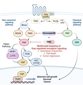
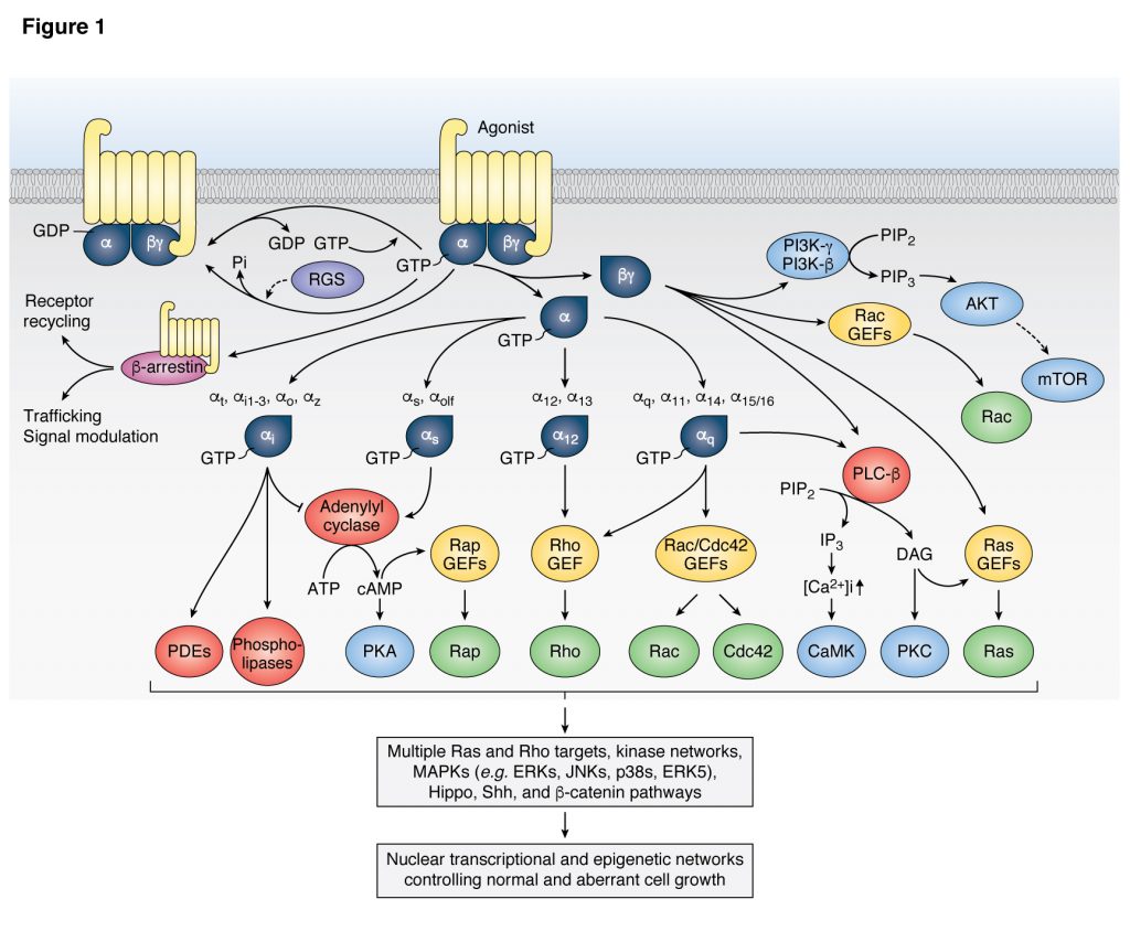
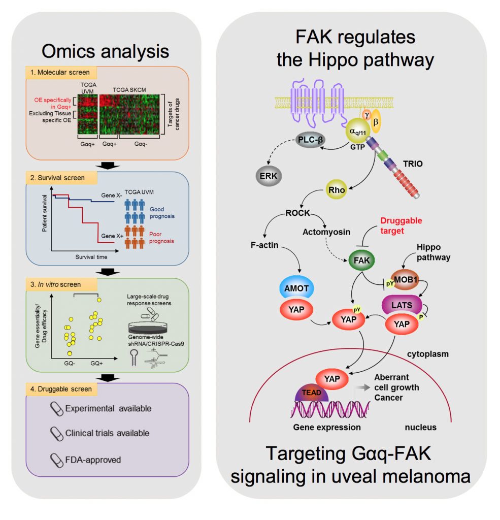
Uveal melanoma (UM) is the most common primary cancer of the eye in adults, and is the second most common melanoma subtype after skin cutaneous melanoma (SKCM). UM is diagnosed in about 2,500 patients each year in the United States alone, nearly 50% of which will die from liver metastasis within 5-10 years after diagnosis, independent of the successful treatment of the primary lesions. Activating mutations in GNAQ and GNA11 (often referred to as GNAQ oncogenes, which encode GTPase deficient and constitutively active Gαq proteins), were identified in ~93% of UM and 4% of SKCM, respectively, where they act as oncogenic driver oncogenes. These findings and our team’s early discovery that activated mutants of Gαq represent a new class of oncogenes firmly established UM and a subset of SKCM as Gαq-driven human malignancies. Indeed, this provides a clear example of a human malignancy that is initiated by gain of function mutations in Gαq and Gα11 proteins. The best-known downstream signaling event initiated by Gαq involves its ability is to activate phospholipase C (PLC) β and the consequent increased hydrolysis of phosphatidylinositol 4,5-bisphosphate (PIP2) to produce two second messengers: inositol 1,4,5-trisphosphate (IP3) and diacylglycerol (DAG). IP3 raises cytoplasmic Ca2+ levels, which stimulates multiple calcium-regulated pathways and, together with DAG, activates classic protein kinase C (PKC) isoforms. However, the molecular events underlying GNAQ-driven malignancies are not yet defined, thus limiting the ability to develop novel anticancer-targeted therapies. In our recent studies, we found that activating mutation of Gαq can trigger YAP translocation into the nucleus and stimulates YAP-dependent transcription, and that this process is independent from PLCβ stimulation but requires the activation of a Gαq-regulated guanine nucleotide exchange factor, TRIO, and the subsequent activation of the small GTPases RhoA and Rac1 and their associated signaling networks. Gαq activation is sufficient to stimulate YAP in animal cancer models, and knock down of Gαq in uveal melanomas harboring GNAQ mutations reduces the nuclear location of YAP, and YAP-dependent gene transcription. Gαq-TRIO-Rho/Rac signaling circuitry contributes to YAP-dependent growth in uveal melanoma, which is the first described GNAQ/GNA11-initiated human malignancy, and, thus, that YAP may represent a novel therapeutic target for uveal melanoma treatment.

However, there are currently limited option to target YAP therapeutically. Thus, there is an urgent need to understand how Gαq promotes cancer growth in order to develop new targeted (precision) therapeutic options. In a recent study, we used a novel computational framework to shed light on Gαq biology and identify systems vulnerabilities for UM, based on the prediction of synthetic (dosage) lethal gene interactions of Gαq. This novel pipeline integrates data from large multiomics cancer datasets, including UM patient transcriptomes and genomes, with in vitro screens (datasets of gene essentiality and dependence and druggable screens (datasets of drug response screens). The top predicted synthetic lethal gene with GNAQ was PTK2, suggesting the potential benefit of targeting the PTK2 gene product (a non-receptor tyrosine kinase known as FAK) in UM. Remarkably, activation of FAK by Gq-linked receptors was initially reported by our team in the early 90s. This unexpected convergence of computational predictions, biochemical, and genetic information prompted us to focus on the role of this Gαq-tyrosine kinase signaling axis in UM. We found that gene editing or inhibition of FAK reduces UM cell growth and tumor formation in vivo. Furthermore, systematic dissection of the underlying mechanisms led us to uncover that FAK promotes YAP activity through direct tyrosine phosphorylation of YAP concomitant with the release of inhibitory core Hippo signaling on YAP. Our studies provided a novel direct link between Gαq-FAK driven tyrosine phosphorylation networks and YAP activation. Based on these findings, the use of FAKi for the treatment of metastatic UM is under current evaluation, as a single agent and as part of a signal-transduction based multimodal therapy.


As part of these studies, we have used unbiased genetic screens to identify new treatment options for UM. We discovered that inhibition of the non-receptor tyrosine kinase FAK and the adaptive activation of the ERK pathway downstream from GNAQ results in UM cell death and regression of liver metastatic lesions. This provided the foundation for the first signal transduction-based multimodal precision therapy in metastatic UM (mUM)(NCT04720417). Our recent large chemogenetic drug screen revealed that targeting FAK and PKC/PKN represents an effective precision-guided combination, which we plan to explore in the clinic shortly.